 An official website of the United States government




We use 3-D bioprinting to create models that mimic human tissues to speed drug discovery and development.
About the 3-D Tissue Bioprinting Program
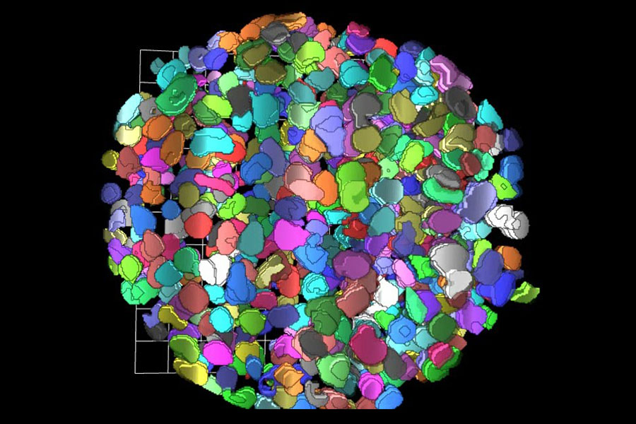
Computed 3-D rendition of the cells in a tumor sphere model after applying a clearing protocol that enables measurements of fluorescence signal from cell nuclei stained with a fluorescent dye, deep inside the tissues.
About 90% of potential new drugs fail in clinical trials because they are not effective or they cause unexpected toxic effects. This failure happens in large part because the cell-based laboratory tests, called in vitro assays, and animal models used during the drug discovery and development process are too simplistic and do not accurately predict what will happen when the drug is given to humans.
There is a critical need for development of in vitro assays that can better predict the effectiveness and toxicity of potential drugs in humans. To address this need, we are applying the techniques of 3-D bioprinting to drug discovery and development. Through the process of 3-D bioprinting, researchers are developing tissue models that mimic the 3-D structure and organization of cells in the tissues of living organisms in microplate format for screening.
The Technology
Recent advances in the fields of engineering, biomaterials science, stem cell biology, physics and medicine have enabled the field of 3-D bioprinting of living tissues. Living cells and scaffolding materials can now be combined into complex 3-D functional tissues produced on microplates (tissue-in-a-well), which are used in preclinical drug testing. These tissues are created using either induced pluripotent stem cells (iPSCs) or primary cells taken directly from living tissue.
The goal of creating 3-D bioprinted human-like tissues in microplate format for screening is to provide physiological and pharmacological data that predict the effects of drugs better than data from traditional studies using 2-D models.
The Applications
At NCATS, the 3-D Tissue Bioprinting team is developing normal and disease tissue models for drug discovery and development. The team has created the infrastructure necessary to enable screening with 3-D bioprinting of tissue-in-a-well models.
The 3-D Tissue Bioprinting program builds on internal expertise in assay development for drug screening, automation technologies, stem cell biology, tissue engineering and advanced high-content microscopy. Learn more about the goals of the program.
The Collaborators
We are collaborating with academic investigators in the basic and clinical research communities who provide the disease and tissue physiology expertise necessary to develop and validate bioprinted tissues. These collaborators might also provide critical materials needed to study drug response, such as patient-derived iPSCs.
We also work with private-sector partners who provide technology and engineering solutions to enable this innovative work. The center is interested in establishing new collaborations with pharmaceutical and biotechnology companies to enable benchmarking and establish translational capacity for these tissue-in-a-well assays so that they can be integrated into the decision making process during the drug discovery and development pipeline.
3-D Bioprinting News
A New 3-D Skin Tissue Model Helps Identify Possible New Treatments for Herpes Simplex Virus
January 27, 2026 - NCATS News
- 3-D Tissue Bioprinting
The 3-D Tissue Bioprinting Program created a 3-D tissue model that mimics human skin to support an infectious disease study focused on identifying potential drug treatments for herpes simplex virus.